461-M AM
Modeling the Relationships between Cortical Surface Reconstruction Methods
Department of Mathematics, Florida State University, Tallahassee, Florida, U.S.A. 32306-4510
Methods: We have created hemisphere cortical surface representations of the GM, WM and the mid-surface using magnetic resonance imaging (MRI) data from 15 different subjects. Surfaces were created using a number of freeware packages, such as BV, CT and FS. Surface characteristics including volume enclosed by the surface and surface area were used as the basis for developing a model to relate results across methods.
Results and Discussion: There is a linear relationship that can be observed between surface area and volume. Furthermore, there is a shift from BV values up and to the right to get comparable FS values. Results for the left hemispheres (LH) and right hemispheres (RH) are shown in Figure 1. We have developed equations from BV to FS values for surface area and volume. Results using other cortical reconstruction packages are also presented.
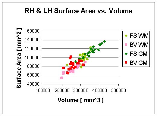
Figure 1: RH and LH surface area versus volume results for BV and FS. A linear trend is observed for hemisphere surface area when plotted against respective volume. There is also an upward and right shift in values when comparing BV to FS.
Conclusions: The mathematical models we have developed allow one to convert FS surface area and volume results to BV and vice versa. We have developed similar equations for conversions across other software packages. These results facilitate the comparison and understanding across studies using one surface reconstruction method versus another.
References & Acknowledgments:
[1] Mangin, J. F. et al. Object-based morphometry of the cerebral cortex. IEEE Trans. Medical Imaging, 23: 968-982, 2004.
[2] Van Essen, D.C. et al. An integrated software suite for surface-based analyses of cerebral cortex. J American Medical Informatics Association, 8:443-459, 2001.
[3] Dale, A. M. et al. Cortical surface-based analysis I: segmentation and surface reconstruction. Neuroimage, 9: 179-194, 1999.
[4] Kline, A. D. et al. Comparison of human cortical surface reconstructions from magnetic resonance imaging data. Neuroimage, 26, Supp. 1: Abstract 631, 2005.
This work is supported in part by NSF grant DMS-0101329, NIH grant P20 EB02013 and a FSU Howard Hughes Fellowship in Mathematical and Computational Biology. We would like to thank Dr. David Rottenberg, Departments of Radiology and Neurology, University of Minnesota for providing the MRI data.