| 11th Annual OHBM Meeting | ||
|
| ||
| Abstract Number: 631 |
Submitted By: Monica K. Hurdal | |
| Last Modified: 11 Jan 05 | ||
1Department of Mathematics, Florida State University, Tallahassee, FL, U.S.A. 32306-4510, 2Department of Radiology, University of Minnesota, Minneapolis, MN, U.S.A. 55455, 3Department of Neurology, University of Minnesota, Minneapolis, MN, U.S.A. 55455, 4Rotman Research Institute, Baycrest Centre for Geriatric Care, Toronto, ONT, Canada M6A 2E1 |
| Objective:
Software which is readily available to the neuroscience community can be
used to reconstruct cortical surfaces from magnetic resonance imaging
(MRI) data. Topologically correct cortical surfaces representing a
white matter (WM) surface (which occurs at the white matter/gray matter
interface) and a gray matter (GM) surface (which occurs at the gray
matter/cerebrospinal fluid interface) can be created. Using these
surfaces, we present an approach for producing a cortical mantle volume
representing the gray matter. We then use this cortical mantle volume
to restrict the analysis of functional MRI (fMRI) data to the gray matter.
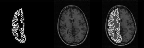
Methods: The FreeSurfer software [1] is used to produce topologically correct WM and GM surfaces from 16 subjects. By default, FreeSurfer translates a surface so the center of the MRI volume is located at the origin. We translate the surface back to "native" volume space so the surface coordinates are located within the original (i.e. native) MRI dimensions. The vertex locations of the triangles which compose the surface are then converted to voxel locations in a manner that ensures no voxel gaps, giving a voxel representation of the cortical surface. A region growing routine is used to identify all voxels enclosed within the WM surface voxel representation and also the GM surface voxel representation. Any voxels inside this enclosed WM voxel region are removed from the enclosed GM voxel region. The result is a voxel mask that represents voxels which belong to the GM surface, voxels which belong to the WM surface and voxels which belong in between the GM and WM surfaces. We call this voxel mask the cortical mantle volume mask. Results & Discussion: Figure 1 shows a cross-section of the resulting cortical mantle volume imposed on an MRI slice. We are using these masks to restrict fMRI analysis to the cortical mantle. FSL [2] is being used to perform GLM analysis on a block design parametric static force BOLD fMRI dataset [3] of the 16 subjects. The SPMs from two single runs for each subject are masked by the cortical mantle mask. Correlation between the two SPMs generated for each single run are computed using the unmasked SPMs and also using the cortical mantle masked SPMs. Conclusions: Construction of a cortical mantle mask permits restricting analysis of fMRI data to the cortical mantle. There have been a number of hypotheses regarding the possibility of improved functional localization results using cortical surfaces. This research represents an attempt at determining whether fMRI analysis that is restricted to the cortical mantle plays a significant role in these hypotheses. References & Acknowledgements: [1] Fischl, B. et al. Cortical surface-based analysis II: inflation, flattening, and a surface-based coordinate system. NeuroImage 9:179-194, 1999. [2] Smith, S. et al. Advances in functional and structural MR image analysis and implementation as FSL. NeuroImage, 23:208-219, 2004. [3] LaConte, S. et al. The evaluation of preprocessing choices in single-subject BOLD fMRI using NPAIRS performance metrics. NeuroImage, 18:10-27, 2003. This work is supported in part by NSF grant DMS-0101329 and NIH grant P20 EB02013. |
 |